Research to better understand the process of arterial overgrowth
WUT researchers’ idea will help to estimate the risk and rate of arterial overgrowth and the risk of haemolysis in coronary artery disease. The project assumes the creation of three-dimensional models based on CT data, reflecting the geometries of the arteries.
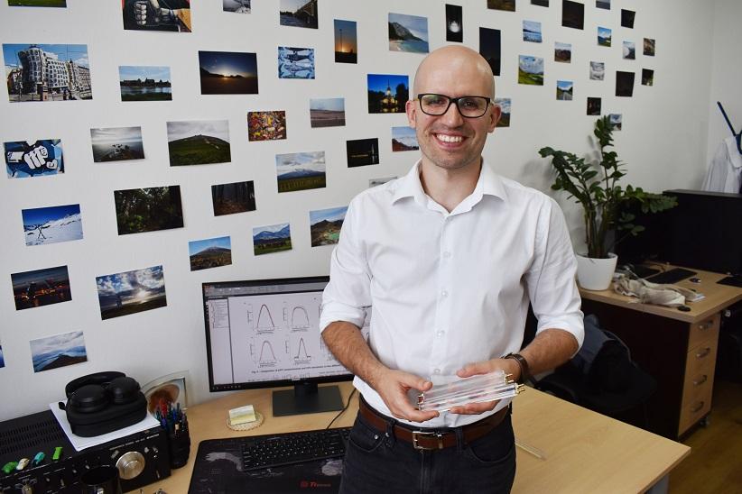
“We have been cooperating with cardiac surgeons from the Medical University of Silesia for several years. They were an inspiration behind our interest in the coronary arteries and the process of haemolysis, which is the death of red blood cells, facilitated by narrowing in this type of artery,” says Krzysztof Wojtas, PhD, from the Faculty of Chemical and Process Engineering at the Warsaw University of Technology.
One of the main medical procedures used to treat vascular obstruction is percutaneous coronary intervention with stents. Stents dilate the arteries and improve blood flow. Although it is a common procedure, the scientific literature lacks a precise theoretical description of the flow and associated phenomena, with experimental verification after a successful surgical procedure. This is due to the fairly recent development of modern computer and research methods allowing scientists to study physiological systems on a micro-scale. So far, they have only been able to guess the nature of the phenomena under analysis.
WUT scientists became interested not only in haemolysis but also in stent overgrowth.
“When we introduce a foreign body into the human body, we cause inflammation. Secondary stent overgrowth occurs on a biochemical as well as a mechanical basis,” explains Dr Wojtas.
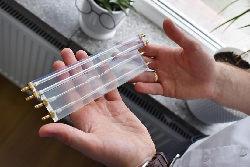
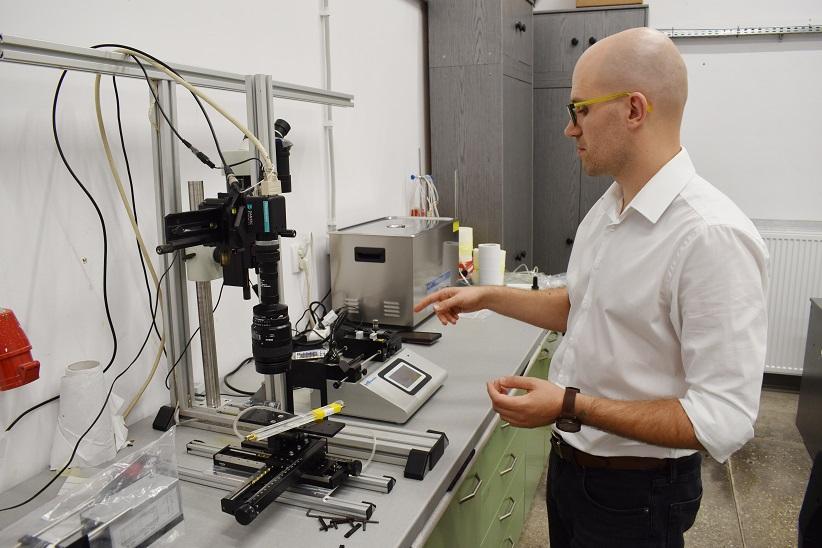
The project assessed different states of the patients' arteries: stenosed before stent insertion, after surgery, and overgrowing at the stent insertion spot. The researchers based their theoretical model on computational fluid mechanics supplemented by population balance, among other things, which facilitated tracking even single red blood cells in the calculations. They also created transparent 3D models of the studied artery. Based on the CT images, they printed 3D models of the patient's artery. They conducted laser velocity measurements using those models. This measurement inside the artery (artery model) served as an experimental verification of their calculations. What does such a laser velocity measurement look like?
“In simple terms, the laser illuminates a given section of the interior of our model, into which we introduce small particles. It takes two pictures with the help of the camera in a noticeably brief time interval (even less than a millisecond). In the pictures, we can see how many pixels the particles have moved between the two shots. This is how we know at what speed they have moved,” explains Dr Wojtas.
A transparent model
During the project implementation, the scientists faced an unexpected challenge. It turned out that the first printouts of the artery model were dull and yellowed, which conflicted with the requirement of transparency and colourlessness of the prints for proper measurements.
“To prepare a print correctly using resins (Low Force Stereolithography technology), they need to be ultraviolet cured and heated. However, overexposure and heat treatment can lead to yellowing. We therefore had to select these parameters by trial and error, as the suggested parameters did not meet our requirements. On the other hand, the tarnish remaining on the outer walls was removed manually with fine-grained sandpaper. We are still looking for a more effective solution,” says Dr Wojtas.
Artificial blood
A mixture of water and glycerine is most commonly mentioned blood substitute in the literature. In the case of the WUT research, where viscosity and fluid velocity are important for low shear rates (in simple terms, low velocities), the WUT scientists added xanthan gum to this mixture to make its behaviour even more bloodlike.
“We do not use real blood to study flow velocity, because of bioethical requirements and cost, and practical problems, as well - nothing would be visible in laser measurements. We do, however, have plans to study haemolysis in this way. The preparations are underway and are complicated, as we need to evaluate the toxicity of the material from which we made the 3D model, among others, to ensure that it will not affect the number of blood cells in the pumped blood,” says Dr Wojtas.
The research initiated under the IDUB project has enabled the WUT researchers to establish cooperation with other centres: the Military Institute of Medicine in Warsaw and the Medical University of Warsaw.
“Through this, we will be able to apply our findings more widely in the future - for example, to analyse the risk of haemolysis in the heart affected by paravalvular leak (a relatively common pathology after valvular replacement surgery) and to analyse phenomena occurring in the carotid and cerebral arteries,” says Dr Wojtas.
The project “Methods for estimating the risk of primary and secondary arterial stenosis associated with coronary artery disease using computational fluid dynamics, flow measurement imaging techniques and 3D printing.” was funded by a research grant under the “Excellence Initiative - Research University” programme, implemented at the Warsaw University of Technology.
Research team:
Krzysztof Wojtas, PhD; Krystian Jędrzejczak, MSc; Arkadiusz Antonowicz, MSc; Michał Wojasiński, PhD; Michał Kozłowski, Doctor of Medical Sciences.


